Watson & Crick’s Structure of DNA Paper in 28 Tweets

Some of you Twitter monsters like myself may have cheered with more of less enthusiasm the change in Twitter policy that the limit of characters per tweet is now doubled to 280 characters as opposed to the previous 140.

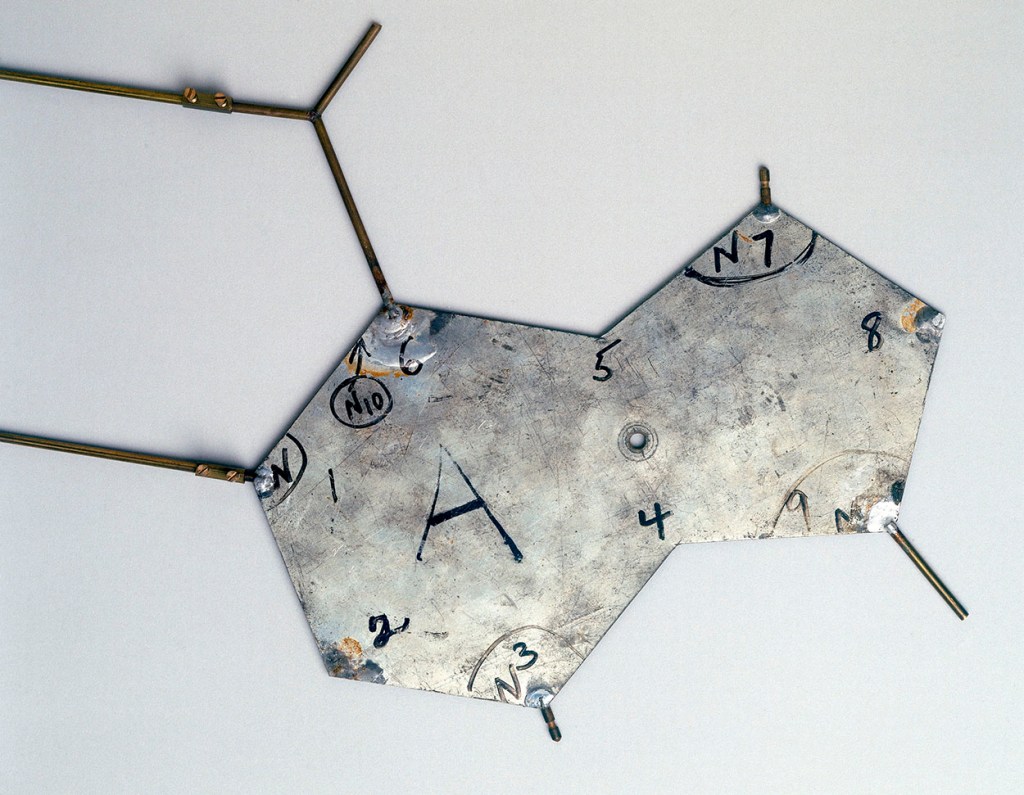
To test how effective this change could be for science, I decided to conduct the experiment of tweeting the 1953 structure of DNA paper published in Nature by Watson and Crick. The only rule I imposed myself was that the chunks I tweeted had to be paragraphs or complete sentences. I only tweeted the main text body. In total, it took me 28 tweets, which I now reference below for your perusal.

Probably this is the first time this landmark paper has been ever shared via this medium.

Template from Crick and Watson’s DNA molecular model, 1953.
This aluminium template representing the base thymine (T) is part of Crick and Watson’s model of DNA. Bases are those groups of atoms that make up DNA’s twin strands. The bases in each of the strands combines to spell out the organism’s genetic code. DNA was discovered by Francis Crick (b 1916) and James Dewey Watson (b 1928) whilst working in the Medical Research Council Unit at the Cavendish Laboratory in Cambridge. In 1953 they constructed a molecular model of the complex genetic material deoxyribonucleic acid (DNA). Their analysis of the double helix shape of DNA explained how genetic information could be copied and passed from one generation to the next. They were awarded the Nobel Prize for medicine and physiology in 1962. (Taken from Wikimedia under CC BY-SA 2.0 license)

Tweet #1

WE wish to suggest a structure for the salt of deoxyribose nucleic acid (D.N.A.). This structure has novel features which are of considerable biological interest.

Tweet #2

A structure for nucleic acid has already been proposed by Pauling and Corey1 . They kindly made their manuscript available to us in advance of publication.

Tweet #3

Their model consists of three intertwined chains, with the phosphates near the fibre axis, and the bases on the outside. In our opinion. this structure is unsatisfactory for two reasons:

Tweet #4

(1) We believe that the material which gives the X-ray diagrams is the salt, not the free acid. Without the acidic hydrogen atoms it is not clear what forces would hold the structure together, especially as the negatively charged phosphates near the axis will repel each other.

Tweet #5

(2) Some of the van der Waals distances appear to be too small. Another three-chain structure has also been suggested by Fraser (in the press). In his model the phosphates are on the outside and the bases on the inside, linked together by hydrogen bonds.

Tweet #6

This structure as described is rather ill-defined, and for this reason we shall not comment on it. We wish to put forward a radically different structure for the salt of deoxyribose nucleic acid. This structure has two helical chains each coiled round the same axis (see diagram).

Tweet #7

We have made the usual chemical assumptions. namely, that each chain consists of phosphate diester groups joining ß-D-deoxyribofuranose residues with 3’,5’ linkages. The two chains (but not their bases) are related by a dyad perpendicular to the fibre axis.

Tweet #8

Both chains follow righthanded helices, but owing to the dyad the sequences of the atoms in the two chains run in opposite directions. Each chain loosely resembles Furberg’s2 model No. 1; that is, the bases are on the inside of the helix and the phosphates on the outside.

Tweet #9

The configuration of the sugar and the atoms near it is close to Furberg’s standard configuration’, the sugar being roughly perpendicular to the attached base. There is a residue on each chain every 3-4 A. in the z-direction.

Tweet #10

We have assumed an angle of 36° between adjacent residues in the same chain, so that the structure repeats after 10 residues on each chain, that is, after 34 A.

Tweet #11

The distance of a phosphorus atom from the fibre axis is 10 A. As the phosphates are on the outside, cations have easy access to them.

Tweet #12

The structure is an open one, and its water content is rather high. At lower water contents we would expect the bases to tilt so that the structure could become more compact.

Tweet #13

The novel feature of the structure is the manner in which the two chains are held together by the purine and pyrimidine bases. The planes of the bases are perpendicular to the fibre axis.

Tweet #14

They are joined together in pairs, a single base from one chain being hydrogen-bonded to a single base from the other chain, so that the two lie side by side with identical z-co-ordinates.

Tweet #15

One of the pair must be a purine and the other a pyrimidine for bonding to occur. The hydrogen bonds are made as follows: purine position 1 to pyrimidine position 1; purine position 6 to pyrimidine position 6.

Tweet #16

If it is assumed that the bases only occur in the structure in the most plausible tautomeric forms (that is, with the keto rather than the enol configurations) it is found that only specific pairs of bases can bond together.

Tweet #17

These pairs are: adenine (purine) with thymine (pyrimidine), and guanine (purine) with cytosine (pyrimidine). In other words, if an adenine forms one member of a pair, on either chain, then on these assumptions the other member must be thymine; similarly for guanine and cytosine.

Tweet #18

The sequence of bases on a single chain, does not appear to be restricted in any way. However, if only specific pairs of bases can be formed, it follows that if the sequence of bases on one chain, is given, then the sequence on the other chain is automatically determined.

Tweet #19

It has been found experimentally3,4 that the ratio of the amounts of adenine to thymine, and the ratio of guanine to cytosine, are always very close to unity for deoxyribose nucleic acid.

Tweet #20

It is probably impossible to build this structure with a ribose sugar in place of the deoxyribose, as the extra oxygen atom would make too close a van der Waals contact.

Tweet #21

The previously published X-ray data on deoxyribose nucleic acid are insufficient for a rigorous test of our structure.

Tweet #22

So far as we can tell, it is roughly compatible with the experimental data, but it must be regarded as unproved until it has been checked against more exact results.

Tweet #23

Some of these are given in time following, communications. We were not aware of the details of the results presented there when we devised our structure, which rests mainly though not entirely on published experimental data and stereo-chemical arguments.

Tweet #24

It has not escaped our notice that the specific pairing we have postulated immediately suggests a possible copying mechanism for the genetic material.

Tweet #25

Full details of the structure, including the conditions assumed in building it, together with a set of co-ordinates for the atoms, will be published elsewhere.

Tweet #26

We are much indebted to Dr. Jerry Donohue for constant advice and criticism, especially on interatomic distances.

Tweet #27

We have also been stimulated by a knowledge of the general nature of the unpublished experimental results and ideas of Dr. M. H. F. Wilkins, Dr. R. E. Franklin and their co-workers at King’s College, London.

Tweet #28

One of us (J.D.W.) has been aided by a fellowship from the National Foundation for Infantile Paralysis.

So, here we go! The whole body text of Watson & Crick’s paper in 28 tweets.

Which one is your favourite? Mine Tweet #24.


Leave a Reply

%d